Excision of Mucocele Using Dental DIODE LASER
Introduction- Mucoceles are known as “mucus filled cavities” usually present in the oral cavity, lacrimal sac, and paranasal sinuses.

Preoperative View (Courtesy – Dr. Sana Farista)
Etiology - Mucus extravasation and mucus retention are the two most frequently occurring primary mechanical obstructive diseases of salivary glands. Formation of mucus extravasation cyst is mainly due to mechanical trauma causing rupture of ductal system of salivary gland and mucin spills into adjacent soft tissues. Mucus retention cyst is formed markedly by obstruction of salivary ductal walls causing dilatation of ducts without spillage of mucin.
Treatment - Although mucoceles may occur literally anywhere in the oral cavity, the most common sites of occurrence are the lower lip. These lesions are soft, fluctuant and closer to the surface. They have a slightly bluish tinge as the mucin shine through. Mucoceles are benign lesions and do not necessarily warrant removal unless their size causes hindrance or they look unaesthetic. There are various treatment aspects available for the management of mucocele like scalpel incision, complete surgical excision, marsupialization, micromarsupialization, intralesional injections of corticosteroids, cryosurgery, laser ablation, sclerosing agent, and electrocautery methods. The conventional treatment includes removal by a surgical excision wherein the entire mucoceles is removed in toto along with any accompanying minor salivary glands to minimize recurrence. One of the more popular methods for mucocele excision is via a diode laser.
The main advantages of soft tissue laser applications are minimal intraoperative bleeding and swelling, minimum postoperative pain, very less surgical time, without any need of suturing after excision because of natural wound dressing due to denatured proteins.
After informed consent, the lesion is infiltrated with local anesthesia, the protective eye wears are worn and then the lesion is excised using soft tissue diode laser with either wavelength of 810nm/980 nm in contact mode or with 450nm wavelength in noncontact or slight contact mode. After the procedure, postoperative instructions are given and analgesic is prescribed on SOS basis. Also, care should be taken to get rid off the cause (sharp cusps or fractured tooth if any) along with the excision to make the treatment outcome more predictive. Here low level laser therapy can also be done over the exposed wound area using 660nm (red light) in noncontact mode to assist in faster healing and to manage postoperative pain.
According to a study by Carlo Fornaini et al 2016, blue light dental lasers provide minimal discomfort to patients and complete healing occurs within 4-5 days with little traces of carbonization in the histopathological investigation. The use of diode lasers in general have been considered to be an effective and safe alternate to the use of scalpel, another study by M Paglia et al 2015 concluded that dental diode lasers is also seen to minimize relapse of mucocoeles in pediatric patients.

Lesion marked with Diode laser (Courtesy – Dr. Sana Farista)

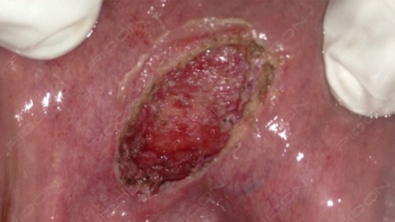
Immediate Postoperative View (Courtesy – Dr. Sana Farista)
Conclusion - Diode dental lasers can be a useful alternative to larger surgical lasers such Erbium and CO2 lasers. Their small size and low cost are distinct advantages. They sterilize the working area, as well as provide coagulation and hemostasis during excisions. Laser application makes it possible to reduce apprehension and fear especially in pediatric and geriatric patients.
References – Fornaini C et al. 450nm diode laser: A new help in oral surgery. World J Clin Cases 2016 September 16; 4(9): 253-257 and M. Paglia etal. Mucocele of the minor salivary glands in an infant: treatment with diode laser. European Journal of Paediatric Dentistry. 2015; 16(2): 139-142.