views

Four Simple Steps for Autograft Bone Harvesting
Autograft bone harvesting, also known as autologous bone grafts, involves the safe extraction of healthy bone from the patient's skeleton. Autograft bones can retain the essential remodeling and regeneration properties of healthy bone. These autologous bones do not carry any disease transmission risks or autoimmune rejections witnessed in using allografts.
Many surgeons prefer to combine autologous bone harvesting procedures with bone graft substitutes to reconstruct bone defects. This procedure is done to maximize the unique properties of autograft bones while minimizing the harvest ratio. Synthetic bone substitutes make the reconstruction process bulky.
Autologous bone grafts are harvested from various skeletal sites, including the fibula and the femur. However, the "gold standard" of bone harvesting is the iliac crest. The reason for this relies on the large mass of harvestable bone in the pelvis.
Autologous bone harvesting techniques
Trapdoor techniques
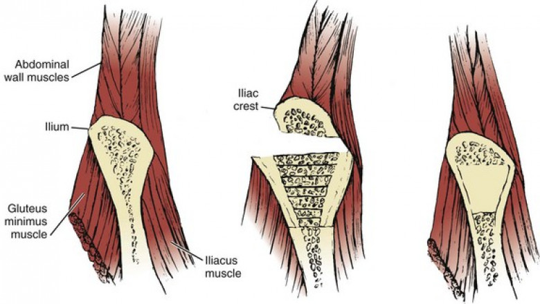
In trapdoor procedures, a bone flap is made in an iliac crest where the subcortical bone graft is extracted before replacing the flap for restoring the cortical anatomy. This is an open method where the cortex is split intentionally to access the cancellous bone.
Splitting techniques
These include any procedures where vertical force is used to make a split or fissure in an iliac crest. It also includes the Wolfe-Kawamoto technique.
Window techniques
In window techniques, a segment of the bone is removed from the iliac crest and the cortex. This procedure is subcrestal to help retain the iliac crest’s aesthetic symmetry.
Trephine extraction
A bone grinder or a trephine can be used to extract a bone graft. These tools can be used to fill a trephine tube to extract a thin layer of cancellous bone graft. The trephine extraction techniques are categorized among the 'minimally invasive' ICBG harvest.
Four simple steps for bone harvesting
- First, you have to mark out an incision, one centimeter, which is nearly two fingerbreadths anterior to the calcaneal tuberosity prominent points. The safest site for bone harvest should be less-superior and posterior to the peroneal tendons.
- After the incision, use a freer elevator to dissect down bluntly into the lateral calcaneal wall bone.
- Break past the lateral wall using a mallet and punch found in the harvesting kit.
- Use a bone graft harvesting system that runs on a standard hand power system. The anterior build of the reamer lets you drive until you get to the far cortex and signal you once you reach the inner wall.
Why you should use the patient’s bone for autograft
-
It takes less time to perform bone harvesting on a patient where the cancellous bone is needed. The autologous bone will be mostly taken from the proximal tibia. This is done using available reusable bone graft harvesting systems.


