views


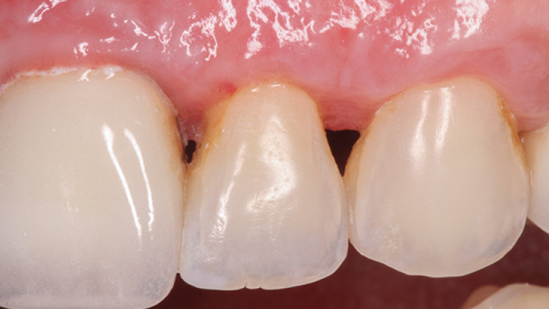
There are several different techniques for treating dental papilla loss. These treatments are graphing the papilla to fill the space, HA injection, and Physiological saline solution. In addition, there are treatments for gingival hyperplasias. This article will discuss each of these options. We will also discuss the risks and benefits of each. Visit Hamilton dental office or read this article. You should feel more informed on the treatment for dental papilla loss.
Graphing the papilla to fill the gap
Graphing the papilla to replace a missing tooth is one method to restore dental apical function and form. It is a highly effective method for restoring papilla function. Researchers from the University of Florida and the University of Michigan studied the effect of graphing the papilla on the position of dental implants. They found a slight correlation between papilla size and contact point. Further, there was no significant correlation between papilla size and distance between bone crests and rehabilitation time.
In one study, a sample of 14 patients with adjacent osseointegrated external hexagon implants was analyzed. The participants ranged from 36 to 78 years, with a standard deviation of 9.3 cm. The Jet index17 and the distance from the dental contact point and bone crest were determined at twenty sites. No significant correlation was found between papilla size and age.
HA injection
An injection of HA gel can be a good treatment for dental papilla loss. The researchers at the University of Florida found that patients who received 5% HA gel experienced a recovery after six months of treatment. One study injected HA into ten patients. The participants were two men and eight women, and the total number of papillae was 35. The study was conducted in the anterior regions of the mandible and maxilla.
A preliminary study also examined the degradability of the HA filler. However, no HA filler survived the Masson Trichrome staining process. Therefore, researchers believe that the presence of HA stimulates bone formation and remodeling activity. However, they do not know if HA can reverse dental papilla loss. However, the study has important implications for dental papilla loss treatment.
Physiological saline solution
The efficacy of physiological saline solution in treating dental papilla loss has been tested by conducting a randomized controlled study. Twenty-four participants with 68 short gingival papillae underwent the treatment with either HA or a physiological saline solution. Participants' gingival papillae were measured using clinical photographs before and after the injection. In addition, the area of black triangles was measured using clinical pictures taken before and after treatment. An in vitro study of gingival fibroblast proliferation and migration was also performed.
Several injection procedures were performed on the same study site:
- The control group was injected with physiological saline solution, while the test group received 16 mg/mL HA gel.
- Injections were repeated six weeks after the first treatment. The study results showed that the treatment was effective in preventing dental papilla loss.
- The study's authors recommend continuing the treatment at least once a year.
Gingival hyperplasias
In a case report, a 32-year-old man was referred to a dental faculty at a university due to bleeding gums and gingival hyperplasia. The patient's medical history was otherwise unremarkable, and an intraoral examination revealed marked hyperplasia of the gingivae. Two days later, he was examined for his thyroid-stimulating hormone (TSH) level, which was elevated. This patient was eventually diagnosed with a dental papilla aplasia and received hormone treatment.
This condition causes a painless swelling in the gingivae and is typically found on the incisor, canine, or premolar region. It can be difficult to distinguish from other gingival tumors, which require histopathologic examination. On the other hand, fibrous hyperplasias are benign, while acanthomatous epulis is malignant and may invade bone. Fortunately, both can be treated therapeutically.


