Glioblastoma multiforme (GBM) is a grade IV brain tumour with a short survival rate. To execute precision surgery followed by chemotherapy treatment, physicians and oncologists urgently require automated tools in clinics for brain tumour segmentation (BTS) and survival prediction (SP) of GBM patients. This blog will look at new approaches for automating the SP process created utilizing automated learning and radiomics. Pre-operative raw magnetic resonance is the best research topic imaging (MRI) images, and clinical data from GBM patients are used in automated algorithms. The general procedure for SP is extracted from all SP techniques submitted for the multimodal brain tumour segmentation (BraTS) competition.
Introduction
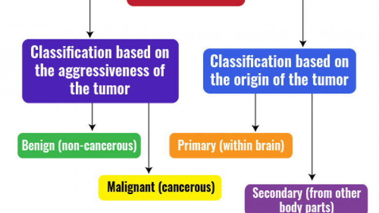
A brain tumour is an uncontrolled proliferation of abnormal cells in the brain. According to a study done in the United States, brain tumours associated with the central nervous system were detected in 23 persons out of every 100,000 people diagnosed each year (CNS). Tumours are classified as benign (noncancerous) or malignant (cancerous) in the medical world based on their aggressiveness and malignancy, as seen in dissertation topics Fig. 1. Primary brain tumours arise from the same brain tissue or adjacent underlying tissues, and primary tumours can be benign or malignant in nature. Malignant tumours that begin elsewhere and quickly spread to brain regions are secondary or metastatic tumours.

Fig. 1 Brain tumours are classified according to their aggressiveness and origin.
Gliomas are the most lethal and severe malignant tumours that arise from the brain’s glial cells. Gliomas account for more than 60% of adult brain tumours. Gliomas include astrocytomas, ependymomas, GBM, medulloblastomas, and oligodendrogliomas. Gliomas are classified into four classes by the World Health Organization (WHO) based on their malignancy, aggressiveness, infiltration, recurrence, and other histology-based features.
Key objectives of the overall survival using pre-operative
Overall survival (OS) prediction offers both benefits and problems because of the abundance of complex and high-dimensional data dissertations in proposal writing services. There is a need to research SP literature based on pre-operative MRI images and clinical data research proposal for PhD. This intends to give readers an overview of the most recent approaches for predicting the survival time of GBM patients. The focus is solely on the BraTS 2020 dataset because it contains the greatest number of cases.
- To investigate the difficulties in using pre-operative MRI scans and clinical data to do automated OS prediction.
- Using the BraTS dataset gives a general workflow of OS prediction algorithms for GBM patients.
- To better understand the assessment measures used to compare the performance of automated OS prediction systems.
- To supply readers and young researchers with useful information regarding BTS and OS prediction using pre-operative MRI images.

Fig-2 Research directions for BTS and OS prediction in the future
Magnetic resonance image analysis for brain tumour treatment planning
Structural MRI is frequently employed in brain tumour research due to its non-invasiveness and higher soft-tissue resolution. Due to imaging artefacts and problems associated with various tumour sub-regions, a single structural MRI is insufficient to separate all tumour sub-regions. Multimodal MRI (mMRI) adds to our understanding of diverse glioma sub-regions. A majority of the tumour is defined by the TC, which is usually excised. Compared to T1-weighted MRI and healthy White Matter (WM) regions in T1ce, areas of T1ce hyperintensity represent the Enhancing tumour. Because T1ce includes both the TC and the ED, the appearance of necrosis (NCR) and non-enhancing tumour (ET) is often less pronounced than in T1. The WT depicts the whole malignant brain area, commonly represented by a circle.
Need for automated techniques
Computer-aided glioma segmentation is critical for overcoming the challenge of radiologists doing manual tumour markings. According to experts, categorical estimations for SP range from 23 to 78 % accurate. At the same time, there are specific challenges, such as picture capture technique variability and the lack of a reliable prognostic model. Biological distinct sub-regions within the tumour such as NCR, NET, ET, and Edema (ED) coexist, which mMRI scans can reveal. Tumour subregions are still difficult to distinguish since they come in various forms and appearances in a PhD literature review.
Challenges in magnetic resonance image analysis
The computer-assisted analysis allows a human specialist to spot the tumour in less time while still preserving the data. Sufficient data and appropriate working processes are required for computerized analysis. The low signal-to-noise ratio (SNR) and abnormalities in raw MRI pictures are caused by radiofrequency emissions created by the thermal mobility of ions in the patient’s body and the coils and electronic circuits in the MRI scanner. Remaining to signal-dependent data biases, image contrasts are diminished due to random fluctuations. Non-uniformity in the intensity of MRI signals is referred to as MRI non-uniformity.
Generic workflow for brain tumour segmentation and survival prediction
Many end-to-end techniques for BTS and SP have been presented in the literature. All of these techniques emphasize their superiority and utility above the others somehow. The BraTS competition is held every year to encourage academics to demonstrate their automated BTS and OS prediction algorithms.
Preprocessing
Data operation algorithms are deep convolutional neural networks (DCNNs). These algorithms need a large amount of data to arrive at relevant findings in the dissertation literature review. Because such large datasets are rarely accessible, preprocessing and data augmentation are needed.
- Min-max normalization
- z-score normalization
- Bias field correction
- Denoising
- Volume cropping
- Intensity clipping
- Spherical coordinate transformation
- Neuromorphic map generation
Post-processing
Several post-processing strategies have been suggested for reducing false positives and enhancing segmentation outcomes. Traditional post-processing approaches, such as threshold- or region-growing methods, use manually determined points to focus on isolated areas or pixels.
- Connected component analysis
- Conditional random field
- Morphological operations
- Relabeling the output label
Fig-3 Future research directions for BTS and OS prediction
Conclusion
The low accuracies found in the literature prompted us to examine several automated approaches and assessment metrics to identify research gaps and other findings linked to GBM patients’ survival prognosis so that future accuracies might be improved. Finally, the report identifies the most interesting future research avenues for improving automated SP approaches’ performance and therapeutic usefulness.
About PhD Assistance
PhD Assistance is a renowned academic guidance provider and has assisted more than 4,500 PhD scholars and 10,500 Masters’s students throughout the globe. We understand your problem in writing the dissertation, and you can seek our help when you find it difficult to keep up with your schedule. In some cases, you would find some parts easy and some parts complex. You can also contact PhD Assistance if you are stuck with your dissertation.